Anatomy of the Arm |
|||||||||||||||||||||
|
Page 04 / 12
Art Book for Learning Human AnatomyThe ForearmANATOMY AND MOVEMENTS The one that is large at the elbow is the ulna. It forms a hinge joint and moves in the bending of the elbow. The other slides as the hinge moves. This second bone is the radius, or turning bone; it is large at the wrist and carries the wrist and hand. Diagonally opposite the thumb, on the ulna, is a hump of bone which is the pivot tor both the radius and also the thumb. Muscles must lie above the joint they move, so that the muscles that bulge the forearm are mainly the flexors and extensors of the wrist and hand. Overlying them and reaching higher up on the arm are the pronators and supinators of the radius. The flexors and pronators (flexor, to flex or bend; pronator, to turn face down, or prone) form the inner mass at the elbow, the extensors and supinators form the outer mass. Between them at the elbow lies the cubital lossa. Both of these masses arise from the condyles of the humerus, or arm bone. These are the tips of the flattened lower end of that bone. From the inner condyle, which is always a landmark, arises the flexor-pronator group. This is a fat softly bulging mass which tapers to the wrist, but shows superficially the pronator teres (round), whose The outer condyle is hidden by its muscular mass when the hand is turned out. This mass is the extensor-supinator group, which bulges higher up, and becomes tendinous half way down. It is dominated by the supinator longus, which rises a third of the way up the arm, widens as far as the elbow, tapers beyond, and loses itself half way down the forearm. In turning, this wedge follows the direction of the thumb, and overlies the condyle when the arm is straight with the forearm. From the back view, the elbow is seen to have three knobs of bone; the two condyles above referred to, and between them the upper end of the ulna, forming the elbow proper, or olecranon. The latter is higher when the arm is straight and lower when it is flexed. The overlying muscular masses meet over halfway down, so that the ulna forms a MASSES The masses of the forearm will be described in connection with those of the arm and shoulder. The ArmANATOMY The bone of the upper arm is the humerus. The part facing the shoulder is rounded and enlarged to form the head, where it joins the shoulder blade. The lower end is flattened out sideways to give attachment to the ulna and radius, forming the condyles. The shaft itself is straight and nearly round, and is entirely covered with muscles except at the condyles. On the flat front side of the condyles, reaching half way up the arm, is placed the broad, flat and short brachialis anticus muscle; and on top of that the thin, high and long biceps, reaching to the shoulder; its upper end flattened as it begins to divide into its two heads. One head passes to the inside of the bone and fastens to the coracoid On the back, behind the flat surface made by the two condyles, arising from the central knob or olecranon, is the triceps (three-headed) muscle. Its outer head begins near the condyle, and occupies the outer and upper part of the back surface of the humerus. The inner head begins near the inner condyle and occupies the inner and lower portion Between biceps and triceps are grooves. The inner condyle sinks into the inner groove below, and it is filled out above by the coraco-brachialis muscle, entering the armpit. The outer condyle sinks into the outer groove below, while midway of the arm the apex of the deltoid muscle sinks into it, overlying the upper ends of both biceps and triceps. 
Bones of the Upper Limb: Muscles of the Upper Limb, front view: 1 Coraco-brachialis. 2 Biceps. 3 Brachialis anticus. 4 Pronator radii teres. 5 Flexors, grouped. 6 Supinator longus. Coraco-brachialis: From coracoid process, to humerus, inner side, half way down. Biceps: Long head from glenoid cavity (under acromion) through groove in head of humerus; short head from coracoid process; to radius. Supination and Pronation of the Forearm, front view: 1 Supinator longus. 2 Pronator radii teres. 3 Flexors, grouped. Supinator Longus: From external condyloid ridge to end of radius. Pronator Radii Teres: From internal condyle and ulna to radius, outer side, halfway down.
Masses of the Arm, Forearm and Wrist Wedging and Interlocking
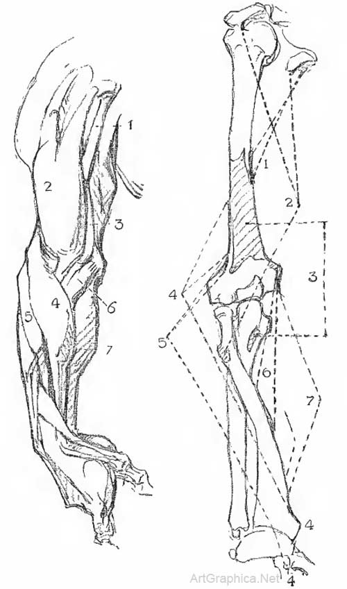
Muscles of the Arm, lateral view (thumb side toward the body) :
I Coraco-brachialis. 1 Biceps. 3 Brachialis anticus. 4 Supinator longus. 5 Extensor carpi radialis longior. 6 Pronator radii teres. 7 Flexors, grouped. Brachialis Anticus: From front of humerus, lower half, to ulna. Extensor Carpi Radialis Longior: From external condyloid ridge to base of index finger. Turning of the Hand on the Forearm AND THE Forearm on the Arm
Muscles of the Upper Limb, outer view:1 Triceps. 2 Supinator longus. 3 Extenso carpi radialis longior. 4 Anconeus. 5 Extensors, grouped. Anconeus: From back of external condyle to olecranon process and shaft ot ulna. EXTENSOR GROUPFrom External Condyle of HumerusExtensor Digitorum Commvmis: From external condyle to second and third phalanges of all fingers. Action: Extends fingers. Extensor Minimi Digiti: From external condyle to second and third phalanges of little finger. Extensor Carpi Ulnaris: From external condyle and back of ulna to base of little finger.
Muscular Mass of Forearm, back view:1 Extensor carpi ulnaris. 2 Extensor communis digitorum. 
Wedging of the Arm into the Forearm, back view
Wedging of Arm into the Forearm AT THE Elbow:1 Biceps. 2 Triceps. 3 Supinator longus. 4 Flexors 5 Extensors.
Muscles of the Arm, inner view:1 Triceps. 2 Biceps. 3 Supinator longus. 4 Flexors, grouped. 5 Pronator teres. FLEXOR GROUPFrom Internal Condyle of HumerusFlexor Carpi Radialis: From internal condyle to first metacarpal. Flexor Carpi Ulnaris: From internal condyle and olecranon to fifth metacarpal, base ot little finger. Flexor Sublimis Digitorum (flexor sublimis perforatus): From inner condyle, ulna and radius to second phalanges of all fingers; perforated to admit passage of profundus tendons.
|
|||||||||||||||||||||
Online Art Books
Human Anatomy Book |


